



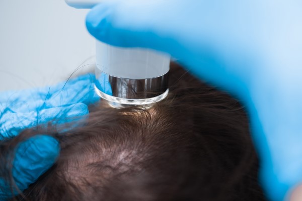
Trichoscopy, also known as dermoscopy or hair and scalp microscopy, is a valuable diagnostic tool in the field of dermatology for the diagnosis of various types of alopecia. This non-invasive technique involves the use of a handheld dermatoscope, which provides magnified visualization of the hair and scalp structures. Trichoscopy allows dermatologists to examine specific features and patterns associated with different types of alopecia, aiding in accurate diagnosis and treatment planning.
When it comes to diagnosing alopecia, trichoscopy offers several advantages over traditional methods. It allows for a close examination of the scalp and hair, providing detailed insights that may not be visible to the naked eye. By identifying specific trichoscopic features, dermatologists can differentiate between various types of alopecia and establish a precise diagnosis.
You may also be interested: Cancer And Hair Loss
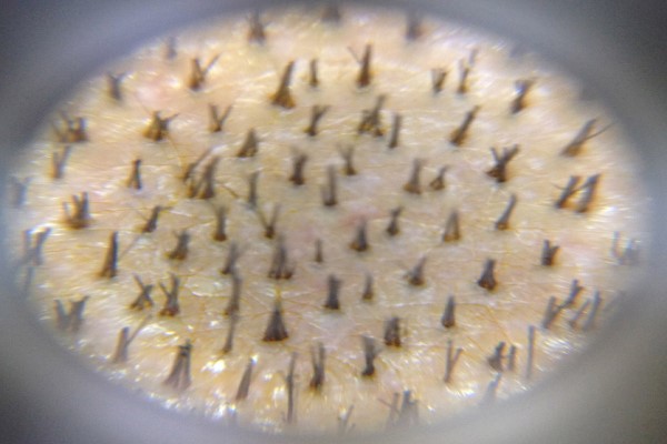
Trichoscopy For Diagnosis of Alopecia
Here are some common types of alopecia and their trichoscopic features:
Androgenetic alopecia (pattern hair loss): Trichoscopy in androgenetic alopecia often reveals characteristic findings such as miniaturized hairs (thin, short, and vellus-like), hair follicle miniaturization, peripilar signs (increased pigmentation around the follicle), and empty follicular openings.
Alopecia areata: Trichoscopy in alopecia areata typically shows “exclamation mark” hairs (broken hairs with a wider proximal end), black dots (exposed or broken hairs in the follicles), yellow dots (dystrophic hairs in the follicles), and short vellus-like hairs. These findings help differentiate it from other forms of alopecia.
Telogen effluvium: Trichoscopy findings in telogen effluvium may include increased telogen hairs (hairs in the resting phase), short regrowing hairs, and tapered hairs. Additionally, the presence of yellow dots and black dots may help differentiate it from other types of alopecia.
Trichotillomania: Trichoscopy in trichotillomania often shows broken hairs of varying lengths, irregularly spaced hairs, and hair shafts with different angles of regrowth. The presence of exclamation mark hairs may suggest the possibility of regrowth.
Traction alopecia: Trichoscopy findings in traction alopecia may include hair shafts with acute bending, broken hairs along the frontal hairline, and a lack of follicular openings in the affected areas.
By carefully examining these trichoscopic features, dermatologists can make accurate diagnoses and tailor appropriate treatment plans for patients with alopecia. Trichoscopy complements clinical evaluation and can help differentiate between different types of alopecia, ensuring that patients receive the most suitable management strategies.
Role of Trichoscopy in Assessing Treatment Response and Monitoring Alopecia Progression
Trichoscopy, also known as dermoscopy or trichoscopic examination, is a non-invasive diagnostic technique used in dermatology to evaluate hair and scalp conditions. It involves the use of a handheld dermatoscope, which provides magnified visualization of the hair and scalp structures. Trichoscopy has proven to be a valuable tool in assessing treatment response and monitoring the progression of various types of alopecia.
When it comes to assessing treatment response, trichoscopy allows dermatologists to objectively evaluate the effectiveness of therapeutic interventions for alopecia. By comparing images taken before and after treatment, they can identify changes in hair density, hair diameter, and the presence of regrowth. Trichoscopic findings can help determine if a treatment is producing positive results, such as increased hair regrowth, reduced hair miniaturization, or decreased inflammation.
Trichoscopy can also aid in monitoring the progression of alopecia over time. By performing regular trichoscopic examinations, dermatologists can observe changes in hair follicles, hair shafts, and the scalp environment. For example, in androgenetic alopecia (pattern hair loss), trichoscopy may reveal characteristic findings such as miniaturized hairs, perifollicular erythema, and follicular units with different hair diameters. Monitoring these features can help assess the rate of hair loss and the effectiveness of interventions.
Furthermore, trichoscopy can be used to differentiate between different types of alopecia. Each type of alopecia has distinct trichoscopic features, allowing dermatologists to make more accurate diagnoses. For instance, trichoscopy can help differentiate between androgenetic alopecia, alopecia areata (patchy hair loss), telogen effluvium (excessive shedding), and other forms of hair loss. This differentiation is crucial for determining the appropriate treatment strategy.
Trichoscopy in Challenging Alopecia Diagnosis
Trichoscopy, also known as dermoscopy or hair and scalp microscopy, can be particularly useful in diagnosing challenging cases of alopecia. In some instances, distinguishing between different types of hair loss can be difficult based on clinical evaluation alone. Trichoscopy provides additional insights by magnifying and examining the hair and scalp structures, allowing dermatologists to identify specific trichoscopic features that aid in the diagnosis of challenging alopecia cases.
Here are some scenarios where trichoscopy can be particularly helpful:
Scarring alopecia vs. non-scarring alopecia: Trichoscopy can help differentiate between scarring alopecia, where irreversible damage to the hair follicles occurs, and non-scarring alopecia, where the hair follicles are still intact. In scarring alopecia, trichoscopy may reveal absence of follicular openings, white dots (indicative of fibrosis), and areas of decreased or absent vascularity. Non-scarring alopecia, on the other hand, may show preserved follicular openings and normal vascularity.
Diffuse alopecia: Trichoscopy can aid in determining the underlying cause of diffuse hair loss, such as telogen effluvium or androgenetic alopecia. In telogen effluvium, trichoscopy may show increased telogen hairs, short regrowing hairs, and tapered hairs. Androgenetic alopecia may be characterized by miniaturized hairs and hair follicle miniaturization.
Cicatricial alopecia subtypes: Trichoscopy can assist in differentiating various subtypes of cicatricial alopecia, such as lichen planopilaris, frontal fibrosing alopecia, and discoid lupus erythematosus. Each subtype may exhibit distinct trichoscopic features, such as perifollicular scaling, perifollicular erythema, follicular hyperkeratosis, and areas of follicular dropout.
Alopecia areata variants: Trichoscopy can aid in diagnosing atypical variants of alopecia areata, such as ophiasis (hair loss along the scalp’s periphery), sisiapho (band-like hair loss), or diffuse alopecia areata. These variants may present unique trichoscopic patterns, such as exclamation mark hairs, yellow dots, or black dots, which can help differentiate them from other forms of alopecia.
By utilizing trichoscopy in challenging alopecia cases, dermatologists can obtain additional information that may not be evident through clinical examination alone. This enhances diagnostic accuracy, leading to appropriate management strategies and improved patient outcomes.
Discovering Various Trichoscopic Elements
Trichoscopy, a valuable technique, enables the identification of four fundamental structures:
- Follicular Openings
- Perifollicular Skin
- Hair Shafts
- Vascular Structures
To ensure a comprehensive assessment, each structure must undergo a thorough examination. Consequently, any report must encompass both normal and abnormal observations. This holistic approach to data interpretation contributes significantly to achieving precise and reliable diagnoses.